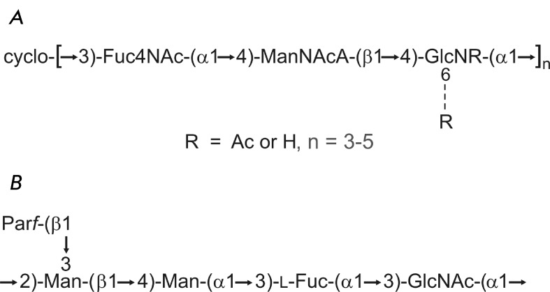
Fig. 3.
Structure of the polysaccharide antigens of Y. pestis (A) and Y. pseudotuberculosis O:1b (B). (A) The cyclic form of the common enterobacterial antigen of Y. pestis [26]. The glucosamine residue is N‑acetylated by ∼50 % and 6-O-acetylated by ∼20 %; n = 4 (major variant), 3 or 5 (minor variants). (B) The pentasaccharide repeating unit of the O-antigen of Y. pseudotuberculosis O:1b [28]. A nonfunctional gene cluster for biosynthesis of this polysaccharide is present in the genome of Y. pestis [29]. Par represents 3,6-dideoxy -D - ribo -hexose (paratose). All monosaccharides have the D configuration; paratose occurs in the furanose form, and the other monosaccharides occur in the pyranose form.

