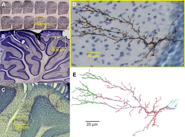
Figure 1.
CF histology. A–D, Rat cerebellar climbing fibers anterogradely labeled with BDA. Yellow boxes indicate regions magnified in subsequent panel. Somata are counterstained with thionine (blue). E, CF digital reconstruction and branch types: primary (purple), tendril (red), retrograde (cyan), and distal (green). Radius thickened 1.5× for clearer visualization.

