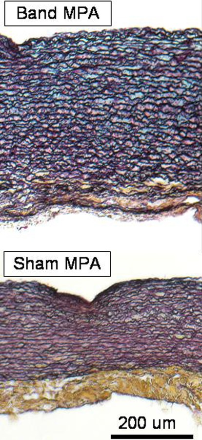
Fig. 4.

Histological images of sham and banded main PAs using Movat's Pentachrome stain (cut along circumference of artery). Collagen fibers (yellow), smooth muscle cells/fibrin (red), ground substance (blue), and nuclei/elastic fibers (black). Scale bar is 200 um.
