
FIGURE 4.
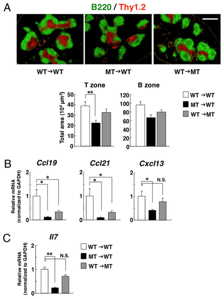
Importance of hematopoietic SIRPα for development of the splenic T cell zone. A, WT or SIRPα MT mice were lethally irradiated and then reconstituted with 5 × 10⁵ BM cells from WT or MT mice for generating WT→WT, WT→MT, or MT→WT chimeras. Eight weeks after transplantation, spleens were harvested, and frozen sections of the spleen from each chimera were stained with mAbs to B220 (green) and to Thy1.2 (red). Scale bar, 500 μm (upper panels). The area for Thy1.2-positive T cell zone or B220-positive B cell zone was measured per each image by the use of Image J software (National Institutes of Health). Data are means ± SE of eight to nine mice per group in two independent experiments (lower panels). Expression of CCL19 (Ccl19), CCL21 (Ccl21) or CXCL13 (Cxcl13) mRNA (B) or IL-7 (Il7) mRNA (C) in the spleen of WT→WT, WT→MT, or MT→WT chimeras was evaluated by quantitative PCR. The level of expression of each mRNA was normalized to that of GAPDH mRNA and presented as fold increase relative to the value for WT→WT chimeras. Data are means ± SE of four mice per group and are representative in two independent experiments. *p < 0.05, **p < 0.01 (one-way ANOVA, followed by the Tukey-Kramer test).