Figure 5.
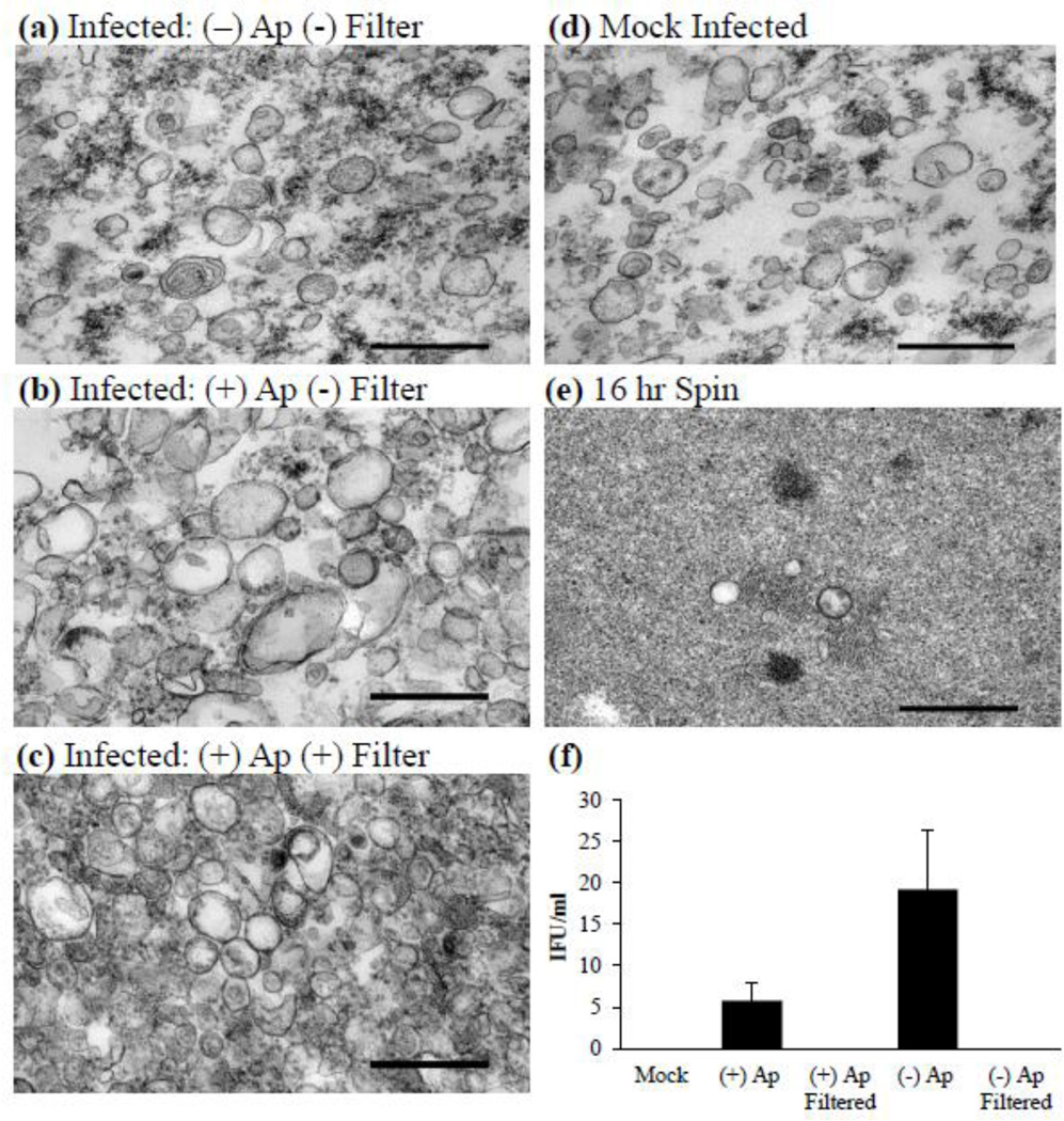
Vesicle purification. (a–e) Representative transmission electron micrographs of various vesicle purifications from infected cultures that are (a) non-ampicillin-exposed, non-filtered; (b) ampicillin-exposed, non-filtered; (c) ampicillin-exposed, filtered; (d) mock infected; or (e) pellet collected from the vesicle isolation supernatant after 16 hr of centrifugation. Experiments performed with the initial fraction (15,300 ×g vesicle supernatant) confirmed that vesicles remain intact after 16 hr of centrifugation. (f) An endpoint assay determining the IFU present in each fraction was conducted to determine possible C. trachomatis elementary body contamination. Values represent mean IFU ± SD from three independent vesicle preparations. Scale bars represent 0.5 µm. Abbreviations: (+) Ap, vesicles isolated from an infection exposed to ampicillin; (−) Ap, vesicles isolated from an infection not exposed to ampicillin.

