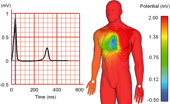
Figure 6.

Simulation of human body-surface ECG, using a biophysically detailed cell electrophysiology model, embedded into an anatomically representative human heart mesh inside a whole-body mesh (containing sub-structures with distinct electrical properties). Left: simulated Lead-I ECG, as measured between the white points indicated on the human mesh (right). The time point of body surface voltage snapshot (on the right) is indicated relative to the ECG by a blue line (on the left). Image courtesy of Nejib Zemzemi, University of Oxford, using techniques developed in (Zemzemi et al., 2011).
