Figure 4.

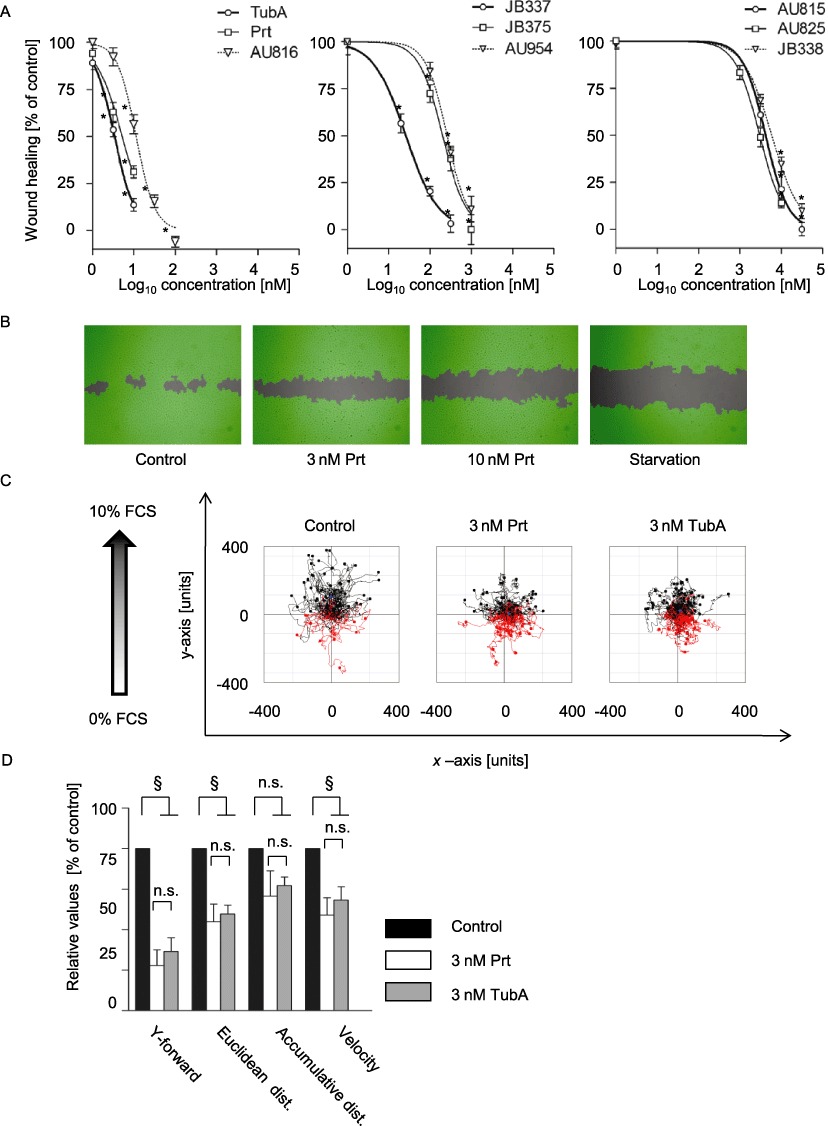
TubA, Prt and Prt derivatives concentration-dependently inhibit endothelial cell migration (A) TubA, Prt and Prt derivatives inhibited wound closure of a scratched HUVEC monolayer with potency similar to the observed effects on proliferation, cell cycle or apoptosis. Data are means ± SEM of three independent experiments, *P < 0.05 vs. untreated controls. (B) Representative images of the wound closure in the absence or presence of pretubulysin or in the absence of growth factors ('starvation’). The cell-free area is grey, the area covered with cells as detected by the imaging software is depicted in green. (C) Representative tracking of the chemotactic movement of endothelial cells in a serum gradient. The starting point of each single cell is placed in the centre of the diagram. Red tracks: cells migrating against the gradient; black tracks: cells migrating along the gradient. In controls, most cells migrated directionally, while cells lost their sense of direction in the presence of Prt or Tub A. (D) Quantitative analysis of the chemotaxis experiments shows reduced parameters of directionality (Y-forward and Euclidean distance), while motility as such (accumulative distance) is not significantly inhibited. Data are means ± SEM of three independent experiments. §Significantly different from controls, P < 0.05; n.s., not significant.
