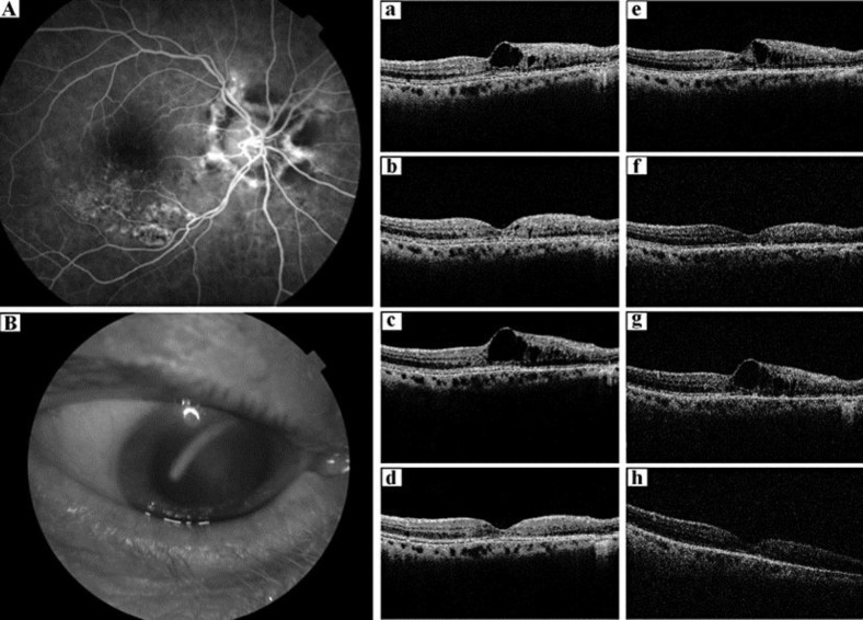
Fig. 1.
FA, slit lamp and macular OCT. A FA shows inferior ME and mild ischemia secondary to inferior temporal vein occlusion with laser scars in the macular area. B Slit lamp examination shows the intravitreal implant just behind the lens with mild cortical cataract. a–h Macular OCT vertical scans. a First exam 3 months after IVT of triamcinolone, cystoids ME on the inferior part of the macula. b 1 month after the first DII with a huge decrease of ME. Persistence of 2 microcysts. c Exam 5 months after the first DII with recurrent ME. d OCT 1 day after the second DII with normalization of the foveal profile. e Recurrence of mild ME 4.5 months after the second DII. f OCT 1 month after the third DII with disappearance of ME but signal deterioration of OCT due to lens opacification. g Exam 4.5 months after the third DII with recurrent ME. h Vanishing of ME, but increase of signal deterioration explaining the bad quality of the examination.

