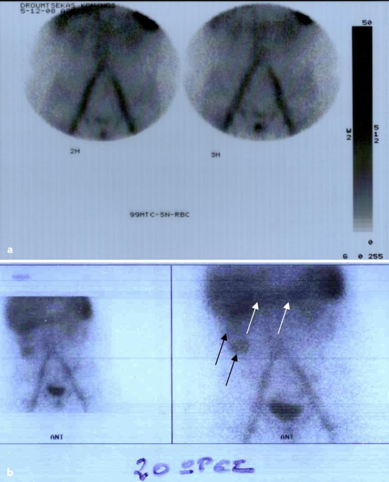
Fig. 3.
Scintigraphy with 99mTc-RBC. a First study period (3.5 h). No abnormal concentrations of the radionucleotide were seen in in the abdominal area (active GI hemorrhage unlikely). b Delayed imaging study (20 h). Radionucleotide concentration in the ascending (black arrows) and transverse colon (white arrows).

