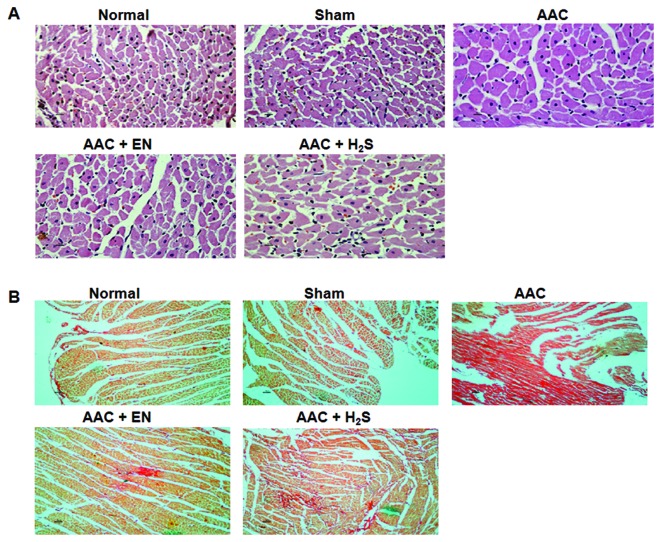
Figure 2.
(A) Cross and longitudinal sections of the left ventricle (LV) stained with hematoxylin and eosin. Original magnification (x200). The minimum size of cardiomyocytes was noted. (B) Collagen deposition. Light micrographs of cardiac fibrosis on LV sections. All sections were stained with picrosirius red stain. Magnification, ×200.

