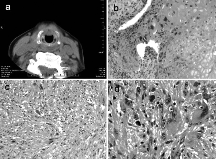
Figure 1.

(a) Axial CT plain scan demonstrating the mass located in the front of the larynx and under the anterior commissure; (b) moderately differentiated SCC, H&E (magnification, ×100); (c) MFH, H&E (magnification, ×100); (d) MFH, H&E (magnification, ×200). SCC, squamous cell carcinoma; MFH, malignant fibrous histiocytoma.
