Membrane repair assay of myofiber transfected with dysferlin-GFP and RFP-MG53.

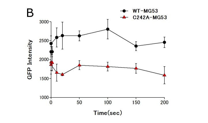
RFP-C242A MG53 perturbed the accumulation of dysferlin at wound sites in the sarcolemma. A. Dysferlin-GFP was simultaneously expressed with RFP-tagged wild-type MG53 or the RFP-C242A-MG53 mutant in mouse skeletal muscle. Arrowheads indicate sites of membrane injury, which were induced with a two-photon laser microscope. Dysferlin-GFP accumulated at the injury site in the presence of RFP-wild-type MG53, but no obvious accumulation of dysferlin-GFP was observed in the presence of the RFP-C242A-MG53 mutant. Scale bar, 10 mm. B.Time course fluorescence intensity (n=3) at wounded sites versus time. For every image taken, the fluorescence intensity of dysferlin-GFP at the site of the damage (circle of 5 mm in diameter) was measured with Zeiss LSM5 Image Examinar software. Data are means ± standard deviation.
