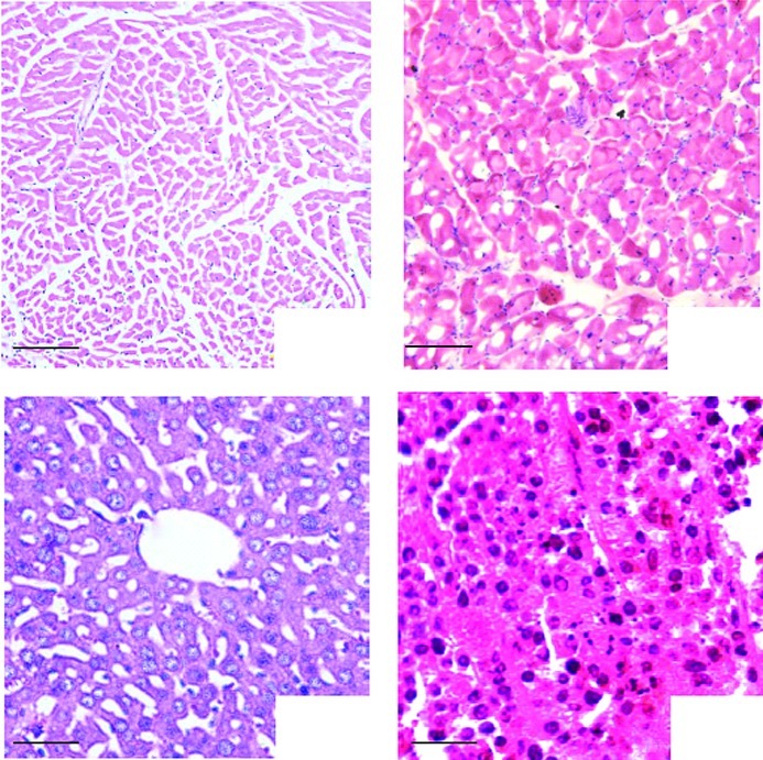
Figure 4.
Histological observations; as mentioned in Materials and methods in the in vivo experimental grouping. (A) Heart, bar, 100 μm, (B) Muscle, bar, 100 μm, (C) Liver, bar, 50 μm, (D) Tumor, bar, 100 μm; H&E staining demonstrated that tissue damage, inflammation and degeneration were not observed in the representative sections.

