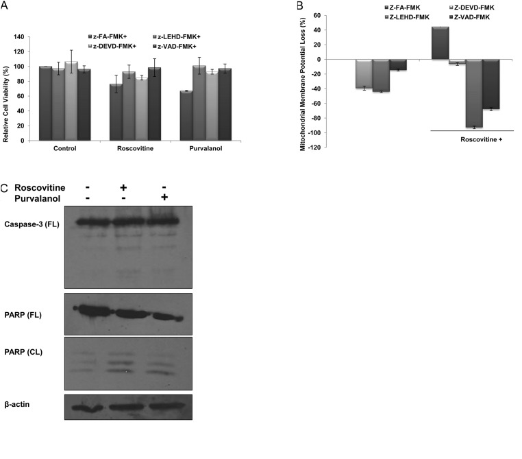
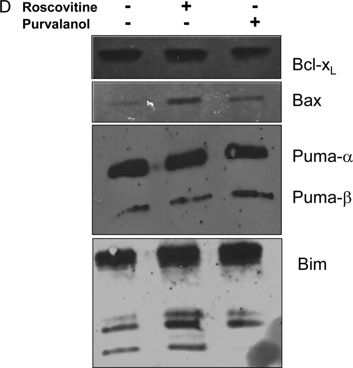
Figure 2.
CDK inhibitors induce apoptosis by activating caspases in Caco-2 cells. (A) MTT cell viability assay was performed to evaluate the role of caspases in drug-induced apoptosis. Following 1 h prior to treatment with caspase-3 (Z-DEVD-FMK), caspase-9 (Z-LEHD-FMK), general caspase (Z-VAD-FMK) and negative caspase (Z-FA-FMK) inhibitors (2.5 μM each), Caco-2 cells were treated with CDK inhibitors roscovitine and purvalanol for 24 h. (B) The disruption of MMP was measured following DiOC6 staining using a fluorometer (Ex = 485 nm; Em = 538 nm). (C) Activation of caspase-3 and PARP cleavage due to apoptotic induction was determined by immunoblotting. Total protein (30 mg) was loaded into each well. β-actin was used as loading control. Columns represent the means ± SD of five replicates from two different culture conditions. (D) Immunoblotting assay was performed to evaluate the modulation of Bcl-2 family members, such as Bcl-XL, Bax, Puma and Bim, following CDK inhibitor treatment in Caco-2 cells.


