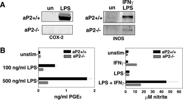
Fig. 4. aP2−/− macrophages show reduced inflammatory activity.
A, deficiency of aP2 diminishes production of proteins from NF-κB-regulated genes. aP2+/+ and aP2−/− macrophages were left unstimulated (un) or incubated for 12 h with 100 ng/ml LPS (left panel) or 100 ng/ml LPS + 10 units/ml murine IFNγ (right panel) before Western blot analysis. B, production of COX-2 and iNOS enzymatic products is reduced by aP2 deficiency. aP2+/+ and aP2−/− macrophage cell lines were stimulated with LPS at the concentrations shown for 24 h, and supernatants were assayed for PGE2 content by ELISA (left panel). Data shown are mean ± S.D. of triplicate determinations. aP2+/+ and aP2−/− macrophage cell lines were stimulated with LPS (500 ng/ml) and/or IFNγ (1 units/ml) for 48 h before measuring nitrite content of supernatants. Data shown are mean ± S.D. of triplicate determinations (right panel).

