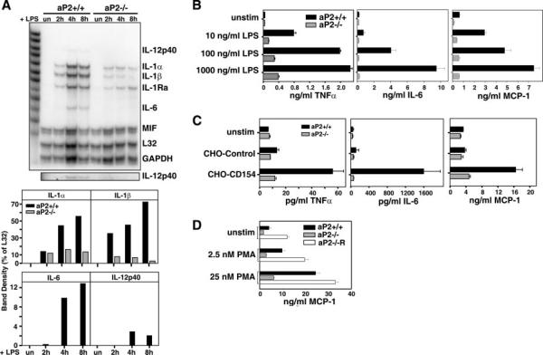
Fig. 5. LPS and CD154-induced expression of inflammatory cytokines are impaired in aP2−/− macrophages.
A, production of inflammatory cytokine RNA is reduced by aP2 deficiency. aP2+/+ and aP2−/− macrophages were stimulated by 1 μg/ml LPS for 0, 2, 4, and 8 h, and cytokine mRNA expression was analyzed by RNase protection assay (top panel). IL-12p40 is shown in a darker exposure in lower panel. Band density of specific RNA is expressed as a percentage of L32 density (bottom panel). GAPDH, glyceraldehyde-3-phosphate dehydrogenase; MIF, migration inhibitory factor. B, LPS induction of inflammatory cytokines and chemokine is reduced by aP2 deficiency. LPS stimulation of cytokines and chemokine production from aP2+/+ and aP2−/− macrophages stimulated for 18 h for IL-6 and MCP-1 and 6 h for tumor necrosis factor α were analyzed by ELISA. C, macrophages were stimulated with CD154 and assayed as in B. Data shown are mean ± S.D. of triplicate determinations. D, aP2 reconstitution into aP2−/− macrophages restores MCP-1 production. Supernatants from aP2+/+ and aP2−/− macrophage cell lines were evaluated for MCP-1 secretion by ELISA after 0 (unstim), 2.5, or 25 nm PMA treatment for 6 h. Data are presented as mean ± S.E. of three determinations.

