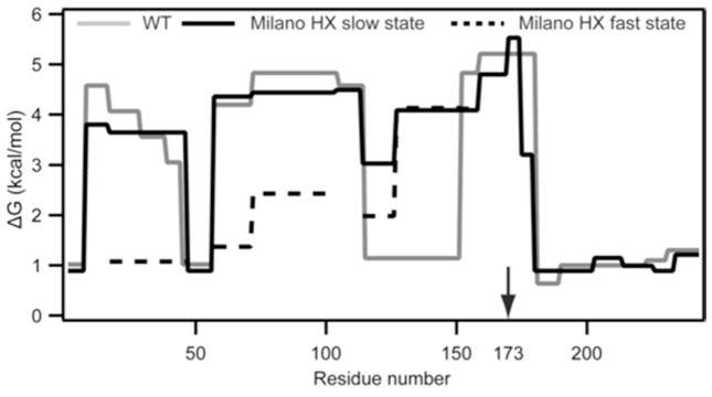
Figure 7.
Summary of the HX-derived secondary structure stabilities for lipid-free apoA-IWT and apoA-IMil. Detailed HX kinetic data (pD 7.3, 5°C) analyzed to obtain Pf values for each peptide are in (Table S2). The corresponding free energies of stabilization are plotted as a function of apoA-I sequence position (apoA-IWT, grey line; apoA-IMil slow and fast HX states are shown with solid and dashed black lines, respectively).

