Figure 4. .
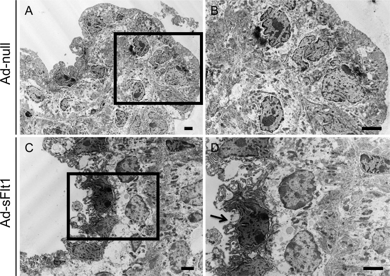
Alterations in ciliary body ultrastructure following VEGF-A neutralization. Ultrastructural analysis of the ciliary body 14 days post-infection illustrates the normal ultrastructure of (A, B) Ad-null animals in comparison to the degeneration of the nonpigmented epithelial layer observed in (C, D) Ad-sFlt1–expressing mice. Arrow indicates the shrunken and nearly nonexistent cytoplasm of a nonpigmented epithelial cell. Scale bar: 2 μm.

