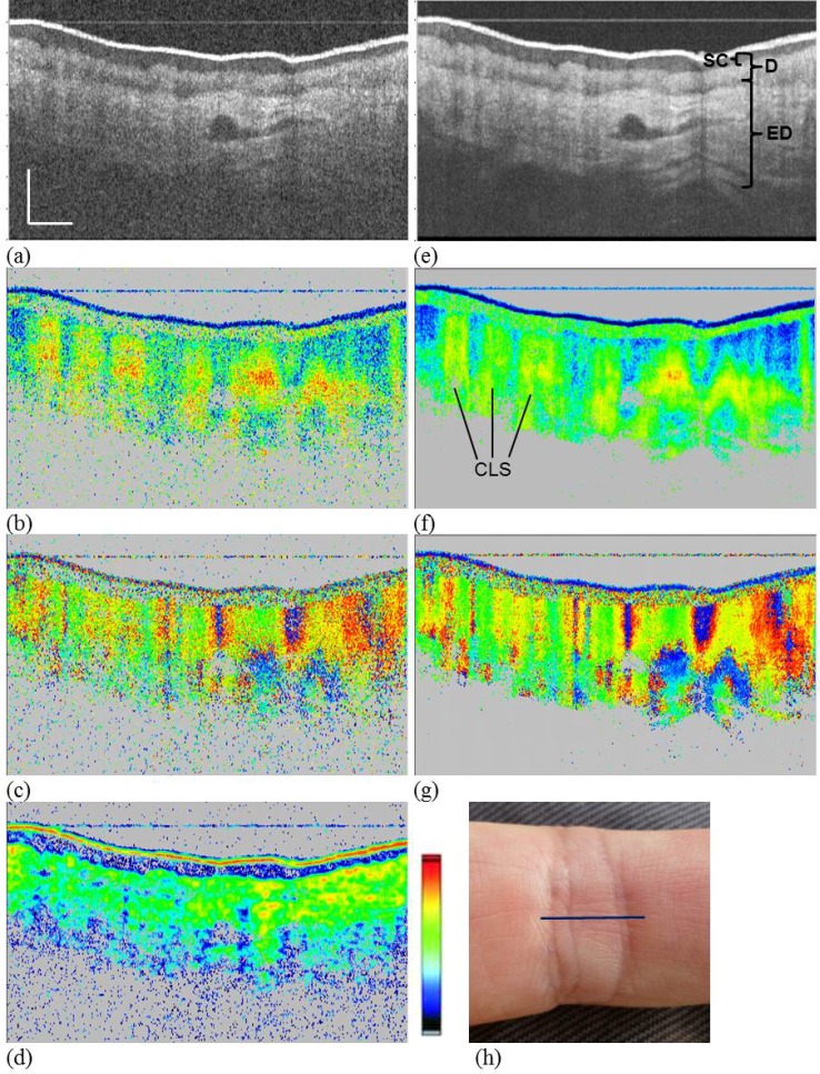
Fig. 4.
PS-OCT images of human skin. Proximal interphalangeal joint of middle finger (PIP) region. (a)–(d) single frame images; (e)–(g) average of 15 frames. (a), (e) reflectivity (log scale); (b), (f) retardation (color scale: 0°–90°); (c), (g) axis orientation (color scale, −90° to +90°); (d) DOPU (color scale, 0–1), 2D DOPU window (12(x) × 6(z) pixels or 55 × 38 µm2); (h) photo of imaged area, line shows approximate B-scan position. Scale bar dimensions: 0.5 mm (x, geometrical distance; z, optical distance). SC, stratum corneum; ED, epidermis; D, dermis; CLS, “column” like structure.

