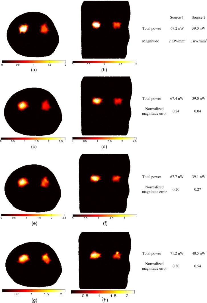
Fig. 3.
BLT reconstruction of two uniform bioluminescence sources localized in the left and right kidneys (4 mm diameters). The “anatomy” is shown in Fig. 1. The actual sources in transverse section (a) and coronal section (b) have magnitudes 2 nW/ mm3 and 1 nW/ mm3 for the left and right source respectively. The reconstructed sources are shown in (c) and (d) using absolute data, (e) and (f) using relative data, and (g) and (h) using imperfect calibrated data. The total powers of the actual and reconstructed sources and the normalized magnitude errors of the reconstructed sources using different reconstruction methods are shown in the figure.

