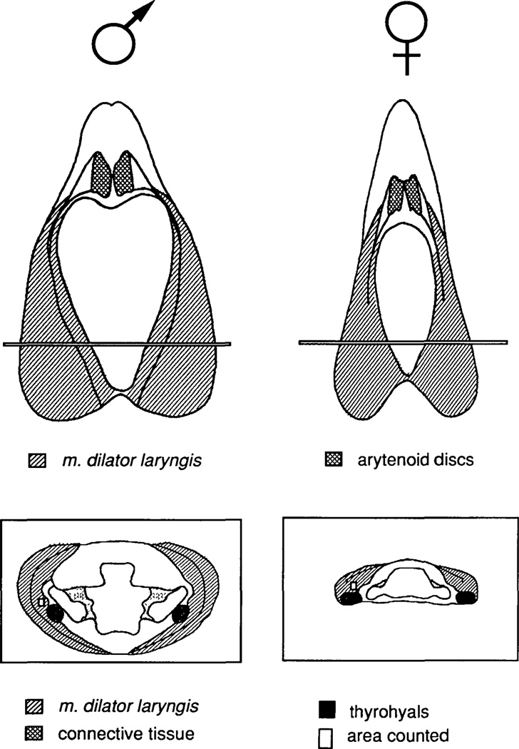
Fig. 1.
Dorsal views (top) and cross sections (bottom) of adult male and female larynges for a typical male and female larynx. For the dorsal views, anterior is up. For the cross sections, dorsal is up. Cross-sections are taken from the anterior-posterior level indicated in the laryngeal dorsal views. Fibers were counted from an area (small rectangle in cross sections) within the inner bipennate muscle just dorsal and lateral to the thyrohyal cartilage. The characteristic shape of the lumen of the larynx at this anterior-posterior level is illustrated in the cross sections.

