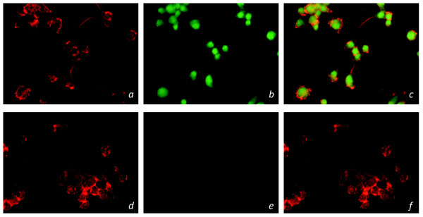
Figure 4.
Immunocytochemical staining of GPR18 receptors in siRNA transfected BV-2 microglia. Immunofluorescent confocal microscopy was conducted using an antibody against the GPR18 C-terminus (1:150; green), phalloidin to label actin (1:40; red). Normal BV-2 microglia with: a) phalloidin b) GPR18 antibody c) phalloidin and GPR18 antibody. GFP+ GPR18 siRNA transfected BV-2 microglia with: d) phalloidin e) GPR18 antibody f) phalloidin and GPR18 antibody.

