
Figure 1.
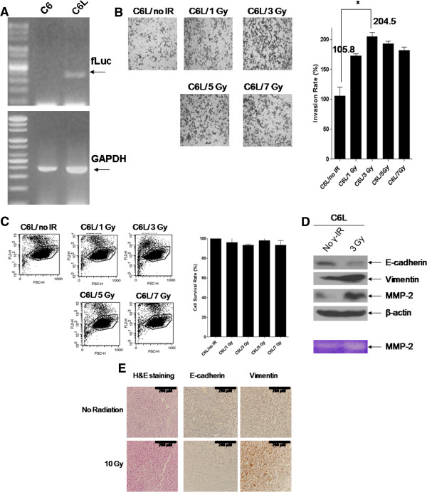
Γ-IR promotes invasion of C6L cells through induction of EMT. (A) C6L, stably transfected cells containing the firefly luciferase gene, was constructed from Rat glioma C6 cells. (B) C6L transfectants were seeded and treated with 1, 3, 5, or 7 Gy of γ-IR, and the invasion assay performed as described in Materials and methods. C6L/no IR represents untreated mock control and C6L/1, 3, 5, or 7 Gy the γ-IR-treated groups. The p values of the C6L/no IR and C6L/3 Gy groups were determined with the t-test. **: p < 0.01 (p value = 0.001). (C) Propidium iodide (PI) uptake analysis. C6L/no IR represents the γ-IR-untreated mock control, and C6L/1, 3, 5, or 7 Gy represents the γ-IR-treated groups. (D) C6L transfectants were treated with 3 Gy of γ-IR, and immunoblot and gelatin zymography analyses performed. No γ-IR represents untreated mock control and 3 Gy signifies the γ-IR–treated groups. (E) Treatment of xenografts with γ-IR also induced EMT, which was detected with IHC (200×). No γ-IR indicates γ-IR-untreated mock control and 10 Gy is the γ-IR-treated group. Γ-IR treatment (10 Gy) was performed as described in Figure 2A, and the tissues collected on day 35 after injection of C6L.