
Figure 4.
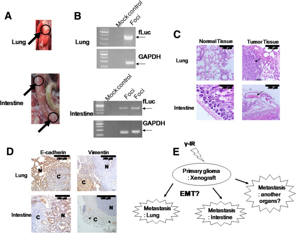
Histological or genetic analysis of metastatic lesions. (A) Mice displaying bioluminescence signals upon imaging were scarified and dissected. Lesions in the lung and intestine were observed (black arrows and circles). (B) The fLuc gene in lesions was detected with PCR. (C) Histological analysis revealed that each lesion displays characteristics typical of cancer tissue (200×). (D) IHC analysis. E-cadherin was expressed in normal tissue, while vimentin was expressed in cancer tissue (N: normal tissue, C: cancer tissue) [200× except to vimentin staining of intestine (100×)]. (E) Scheme of γ-IR – induced metastasis.