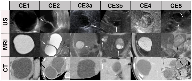
Figure 5. “Worst case” of CT/MR imaging.
The “double line sign”, typical for CE1 is often seen in US (CE1/US, arrow), less reliably in MRI and CT. Daughter cysts and detached endocyst (“water-lily-sign”) is often missed by CTs, but clearly visible in US and MRI (see CE2, CE3a, arrows). Daughter cysts inside a solid cyst matrix are often not recognized by CT (CE3b, arrows). The CE4-specific canalicular structure is often not visible on CT images. These cysts may be misinterpreted as type CE1 cysts, i.e. staged “active” instead of “inactive”. The identification of calcifications is the domain of CT imaging. MRI does not differentiate well between thick hyaline walls and calcifications. US picks up calcifications only when a dorsal echo shadow is produced (see CE5, arrows). MRI: HASTE sequence, CT: post contrast enhanced images.

