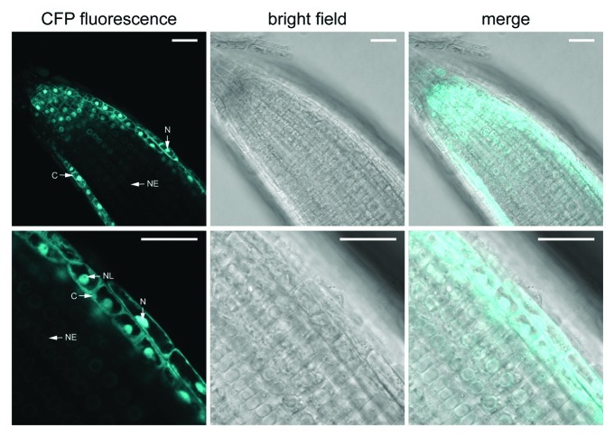
Figure 2. Seh1-CFP subcellular localization in root cells. Confocal images of Seh1-CFP fluorescence in roots of 2-week-old plate-grown seh1–1 seedlings stably expressing Seh1-CFP under control of the double 35S promoter. Scale bars are 25 µm. C, cytoplasm; N, nucleoplasm; NE, nuclear envelope; NL, nucleolus.

An official website of the United States government
Here's how you know
Official websites use .gov
A
.gov website belongs to an official
government organization in the United States.
Secure .gov websites use HTTPS
A lock (
) or https:// means you've safely
connected to the .gov website. Share sensitive
information only on official, secure websites.
