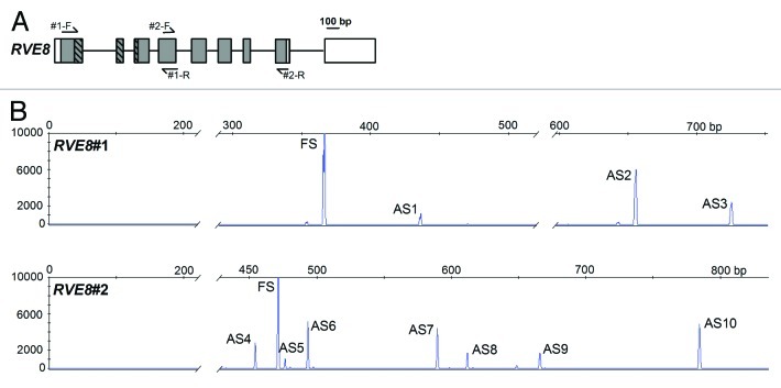
Figure 1. Alternative Splicing of RVE8. (A) RVE8 (At3g09600) gene organization with exons depicted as rectangles, and introns as intervening horizontal lines. Protein coding exons/regions are shaded gray, UTR regions are depicted as clear rectangles. The region of RVE8 encoding the predicted single Myb domain is represented as hatched rectangles. Primer locations are denoted by arrows. Primer sequences are: RVE8#1-F GCTGGACAGAGGAAGAGCAC; RVE8#1-R TGCTCCACGAAGAGTAGCAA; RVE8#2-F GGGATATGCTTCATGGGATG; RVE8#2-R GCTGATTTGTCGCTTGTTGA. (B) Representative HR RT-PCR electropherograms for RVE8. AS isoforms were characterized from pooled RNAs representing plants harvested under both diurnal and constant light conditions at either normal ambient temperature or for plants undergoing cooling. Electropherograms show the size of detected peaks corresponding to RT-PCR products from alternatively spliced transcript variants. The x-axis is the size in bp and the y-axis represents arbitrary scales (relative fluorescent units) to reflect relative abundance of peaks. Individual products are indicated and AS variants are described in more detail in Table 1. FS – fully spliced product; ASn – alternative splicing variant where n is the number to identify the product. AS transcripts were detected on an ABI3730 automatic DNA sequencer along with GeneScan™ 1200 LIZ size standard. Amplicons were accurately sized using GeneMapper software. For more details of Methods see15

An official website of the United States government
Here's how you know
Official websites use .gov
A
.gov website belongs to an official
government organization in the United States.
Secure .gov websites use HTTPS
A lock (
) or https:// means you've safely
connected to the .gov website. Share sensitive
information only on official, secure websites.
