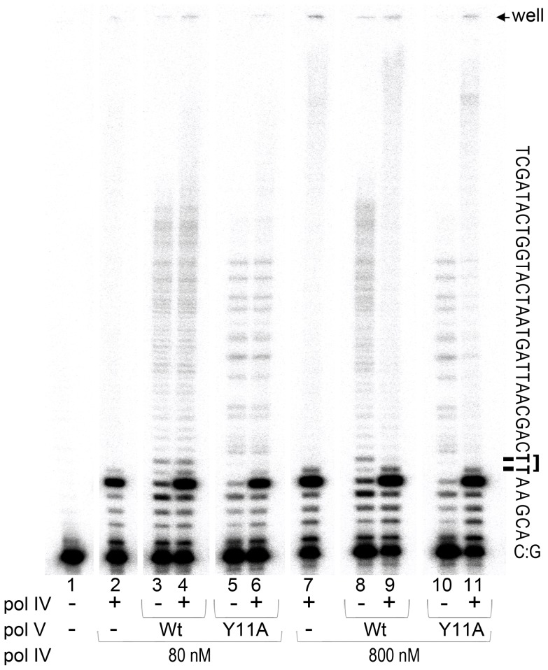
Figure 3. In vitro translesion synthesis past a TT-CPD lesion catalyzed by mixtures of pol IV and pol V.
Translesion DNA synthesis was performed using a circular DNA template with a running-start primer with its 3′ end located 5 bases before the 3′T of the CPD. Primer extension reactions catalyzed by pol IV (80 nM, lane 1 or 800 nM, lane 6), wild-type pol V (80 nM, lanes 2 and 7), pol V (UmuC_Y11A) (60 nM, lanes 4 and 9), or a combination of pol IV (80 nM, lane 3 and 5 or 800 nM, lane 8 and 10) with either wild-type (80 nM, lanes 3 and 8) or polV (UmuC_Y11A) (60 nM, lanes 5 and 10) were performed for 30 sec as described in the Methods section. Part of the template sequence and position of the gel wells and a CPD lesion are indicated to the right of the gel panel. As clearly observed, when present in a 10-fold excess (similar to SOS induced conditions), pol IV inhibits TLS catalyzed by pol V.

