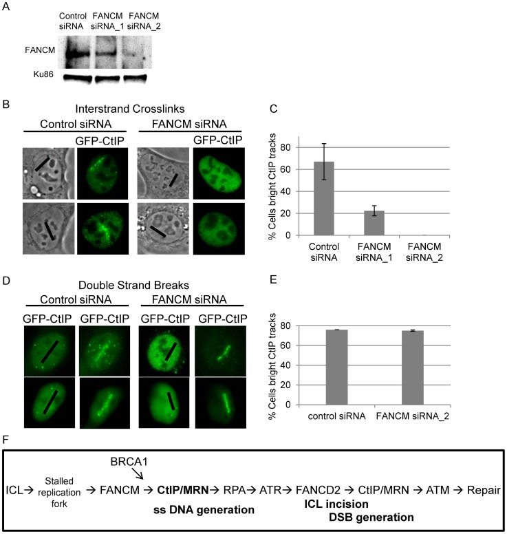
Figure 8. FANCM depletion reduces CtIP accumulation at ICLs but not at DSBs.
(A) Immunoblot of control cells and cells depleted for FANCM. (B) GFP-CtIP in S-phase control and FANCM depleted cells pre-and post-microirradiation with 730 nm light in the presence of 8-MOP. (C) Quantification of cells scored as having visible GFP-CtIP along laser tracks in control or depleted cells. Bars indicate standard deviation between 3 independent experiments. (D) GFP-CtIP in S-phase control and FANCM depleted cells pre- and post-microirradiation with green laser to generate DSBs. (E) Quantification of cells scored as having bright GFP-CtIP along DSB containing laser tracks. (F) Model of CtIP function in replication associated ICL repair. FANCM binds ICL stalled replication fork and remodels fork to enable CtIP and MRN access. BRCA1 ubiquitinates CtIP facilitating its localization to damaged chromatin. CtIP and MRN act to generate ssDNA at a stalled fork. RPA bound single stranded DNA activates ATR. ATR phosphorylates FANCD2 followed by FA core complex ubiquitination of the FANCI-FANCD2 complex. The monoubiquitinated FANCI-FANCD2 complex localizes to the damaged chromatin and facilitates ICL incision and downstream repair events. CtIP and MRN act again to resect ends, activate ATM, facilitate completion of repair by homologous recombination.

