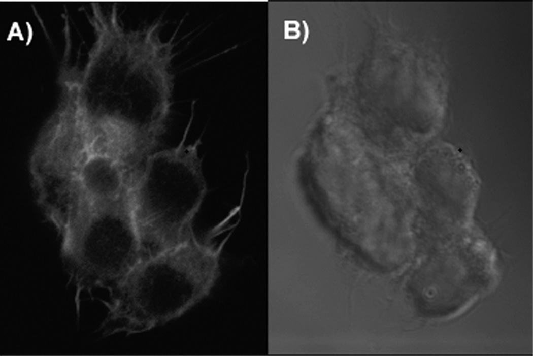
Figure 2.
Lipopeptide inhibitor of Hedgehog pathway fuses with cell membrane spontaneously and saturates intracellular membranes upon prolonged incubation. Confocal laser scanning microscopy of rhodamine red-labeled lipopeptide inhibitor of Hedgehog pathway. A) Peptide fluorescence localizes to intracellular membranes; B), Nomaski image of the cells. For generation of the fluorescent antagonist, Cys-contaning peptide (Pal-CLTYAWHTSFKAL-NH2) was labeled with rhodamine red by reacting cysteine residue with rhodamine maleimide. SK-Mel-2 melanoma cells were exposed to 100 nM solution of the peptide in medium containing Hoechst-33342 for 2 hour and observed under laser scanning confocal microscope (LSCM 510, Zeiss). Rhodamine red fluorescence was excited with 561 nm laser and 575-615 filter was used for emission detection.

