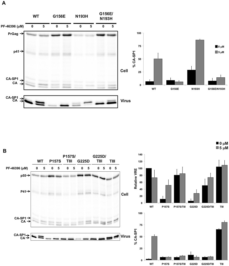
Figure 10. Second-site compensatory changes correct the replication defects exhibited by PF-46396-dependent CA mutants.
[A] Radioimmunoprecipitation analysis of cell- and virion-associated proteins in the absence and presence of 5 µM PF-46396. Analysis performed with WT, CA-G156E, CA-N193H, and CA-G156E/N193H. Positions of Pr55Gag (PrGag), Pr41Gag (p41), CA-SP1, and CA are indicated. Graph on the right shows phosphorimager-based quantification of the % CA-SP1 relative to total CA+ CA-SP1 in virion fraction at the indicated concentration of PF-46396. Error bars denote SD; N = 5. [B] Radioimmunoprecipitation analysis of cell- and virion-associated proteins in the absence and presence of 5 µM PF-46396. Analysis performed with WT, CA-G157S, CA-P157S/SP1-T8I, CA-G225D, CA-G225D/SP1-T8I, and SP1-T8I. Positions of Pr55Gag (PrGag), Pr41Gag (p41), CA-SP1, and CA are indicated. Graphs on the right show phosphorimager-based quantification of relative virus release efficiency (VRE) and % virion CA-SP1, calculated as described in the Fig. 6 legend. Error bars denote SD; N = 3.

