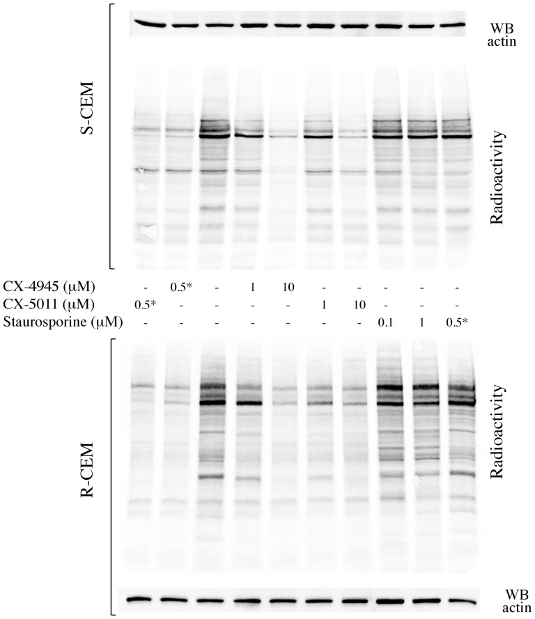
Figure 4. Protein phosphorylation in lysates from cells treated with CX-4945, CX-5011, or staurosporine.
S-CEM (upper panel) or R-CEM (lower panel) were treated with the indicated concentrations of the CX inhibitors or staurosporine for 16 h, then lysed. 5 µg of total proteins were incubated with a radioactive phosphorylation mixture, resolved by SDS-PAGE, blotted, and analyzed by digital autoradiography (radioactivity). WB for actin was used as loading control. Asterisk * denotes samples where the inhibitors were added in vitro during the phosphorylation assay (not administrated to the cells). Representative results of five independent experiments are shown.

