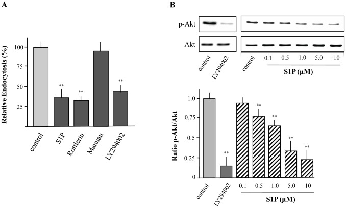
Figure 3. Uptake of FITC-labeled dextran by XS52 cells via macropinocytosis in a PI3K dependent manner.
Cells were preincubated with Rottlerin (3 µM), Mannan (1 mg/mL), and LY294002 (10 µM) for 30 min. Then, cells were incubated with FITC-labeled dextran for 15 min. S1P (5 µM) was used as positive control. Fluorescence intensity of cells was analyzed by flow cytometry and relative endocytosis was calculated. Data are expressed as the mean ± SEM of results from at least three independent experiments. **P < 0.01 indicate a statistically significant difference vs. control experiments (A). Cells were treated with the indicated concentrations of S1P or LY294002 (10 µM) for 15 min followed by the detection of Akt activity using Western blot analysis (B). Values of the densitometric analysis are expressed as x-fold decrease of phosphorylated Akt (p-Akt) formation compared to untreated cells ± SEM from three experiments. **P<0.01 indicates a statistically significant difference versus control (B).

