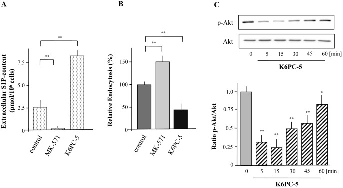
Figure 7. Autocrine modulation of macropinocytosis by XS52 cells via formation and release of S1P.
XS52 cells were cultivated in the presence or absence of MK-571 (15 µM) and K6PC-5 (10 µM) over a time period of 6 h and S1P levels in the extracellular environment was detected (A). Cells were preincubated with MK-571 (15 µM) for 6 h and K6PC-5 (10 µM). Then, cells were incubated with FITC-labeled dextran for 15 min. Fluorescence intensity of cells was analyzed by flow cytometry and relative endocytosis was calculated. Data are expressed as the mean ± SEM of results from at least three independent experiments. **P < 0.01 indicate a statistically significant difference vs. control experiments (B). Cells were treated with 10 µM of K6PC-5 for the indicated time periods followed by the detection of Akt activity (C). Values of the densitometric analysis are expressed as x-fold decrease of p-Akt formation compared to untreated cells ± SEM from three experiments. *P < 0.05 and **P<0.01 indicate a statistically significant difference versus control.

