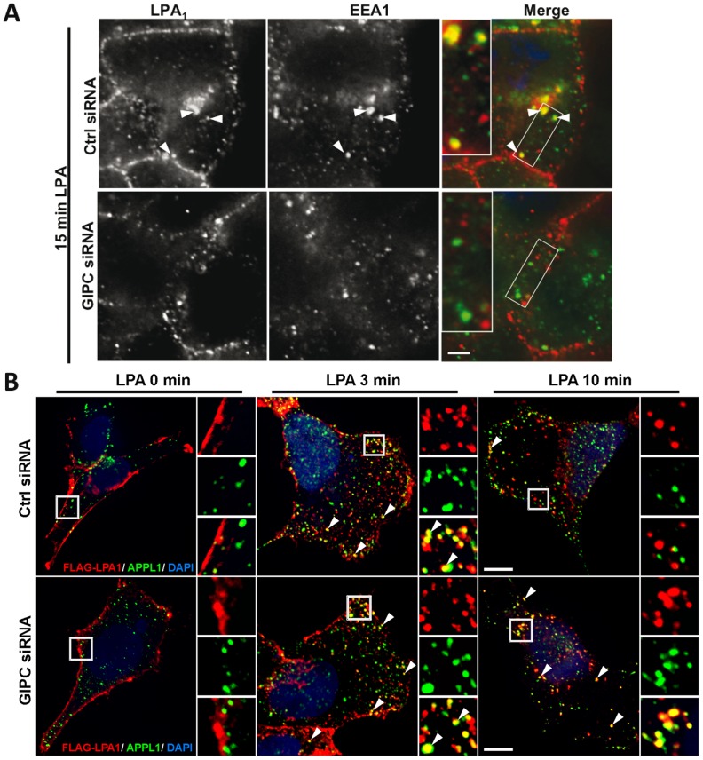
Figure 3. GIPC depletion delays trafficking of LPA1 from APPL1 to early EEA1 endosomes.
A, GIPC depletion inhibits internalization of LPA1 and its trafficking to early endosomes after stimulation with LPA. Upper panel: In HEK-LPA1 cells transfected with control siRNA and stimulated with LPA for 15 min, LPA1 is found in cytoplasmic vesicles where it colocalizes with EEA1 (arrowheads). Lower Panel: In cells transfected with GIPC siRNA fewer vesicles containing LPA1 are present 15 min after LPA stimulation, and less colocalization is seen between LPA1 and EEA1 (compare yellow in right panels). Boxed regions are enlarged (2.2×) in the insets. Images were acquired with a Zeiss AxioImager M1 microscope, and overlap in staining between LPA1 and EEA1 was evaluated using Volocity software. Statistical significance (p value) was determined by t-test. B, Trafficking of LPA1 is delayed in APPL1 endosomes after depletion of GIPC. Left panel: In both GIPC-depleted (GIPC siRNA) and controls (Ctrl siRNA), LPA1 is localized along the plasma membrane after serum starvation (0 min) whereas APPL1 is found in peripheral cytoplasmic vesicles. Middle Panel: In both GIPC depleted and control cells stimulated with LPA for 3 min, LPA1 colocalizes with APPL1 in cytoplasmic vesicles (arrowheads). Right Panel: In controls stimulated with LPA for 10 min, very few LPA1 receptors remain in APPL endosomes (yellow, arrowhead) whereas in GIPC-depleted cells the majority of the receptors are retained in APPL endosomes (yellow, arrowhead). Boxed regions are enlarged (3×) in the insets. HEK-LPA1 cells grown on coverslips were transfected with GIPC or control siRNA. 72 h after transfection cells were serum starved for 4–6 h and subsequently incubated on ice with rabbit (A) or mouse (C) anti-FLAG IgG, shifted to fresh medium containing LPA for the indicated times, then fixed and processed for immunofluorescence using mouse anti-EEA1 IgG (A) or rabbit-anti-APPL1 IgG (C) as in Fig. 2A. Images in “A” were acquired with a Zeiss AxioImage M1 microscope, and those in “C” were acquired with an Ultra View Vox Spinning Disk Confocal. Bar = 10 µm.

