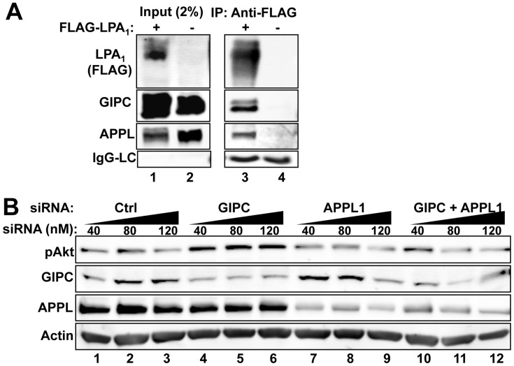
Figure 6. APPL interacts with LPA1 and affects LPA1 mediated Akt signaling.
A, APPL and GIPC co-immunoprecipitate with FLAG- LPA1 (lane 3). HEK cells were co-transfected with HA-APPL and GIPC together with FLAG-LPA1 (lanes 1 and 3) or empty vector (lanes 2 and 4), and cultured in the presence of 10% PBS for 48 h before lysis. IP was carried out as in Fig 1A. Aliquots of cell lysates (input, 2%) were loaded to verify comparable expression levels. B, APPL depletion inhibits Akt activation in HEK-LPA1 cells stimulated with LPA. Depletion of GIPC (lanes 4–6) leads to increased Akt signaling (top panel) compared with controls (lanes 1–3). In contrast, depletion of APPL1 alone (lanes 7–9) or double knockdown of GIPC and APPL (lanes 10–12) results in reduced Akt signaling. HEK-LPA1 cells were transfected with increasing amounts of control (lanes 1–3), GIPC (lanes 4–6) or APPL siRNA (lanes 7–9) or GIPC and APPL siRNA combined (lanes 10–12). Cells were serum starved overnight, stimulated with 5 µM LPA for 15 min, lysed and analyzed by immunoblotting as in Fig. 3D.

