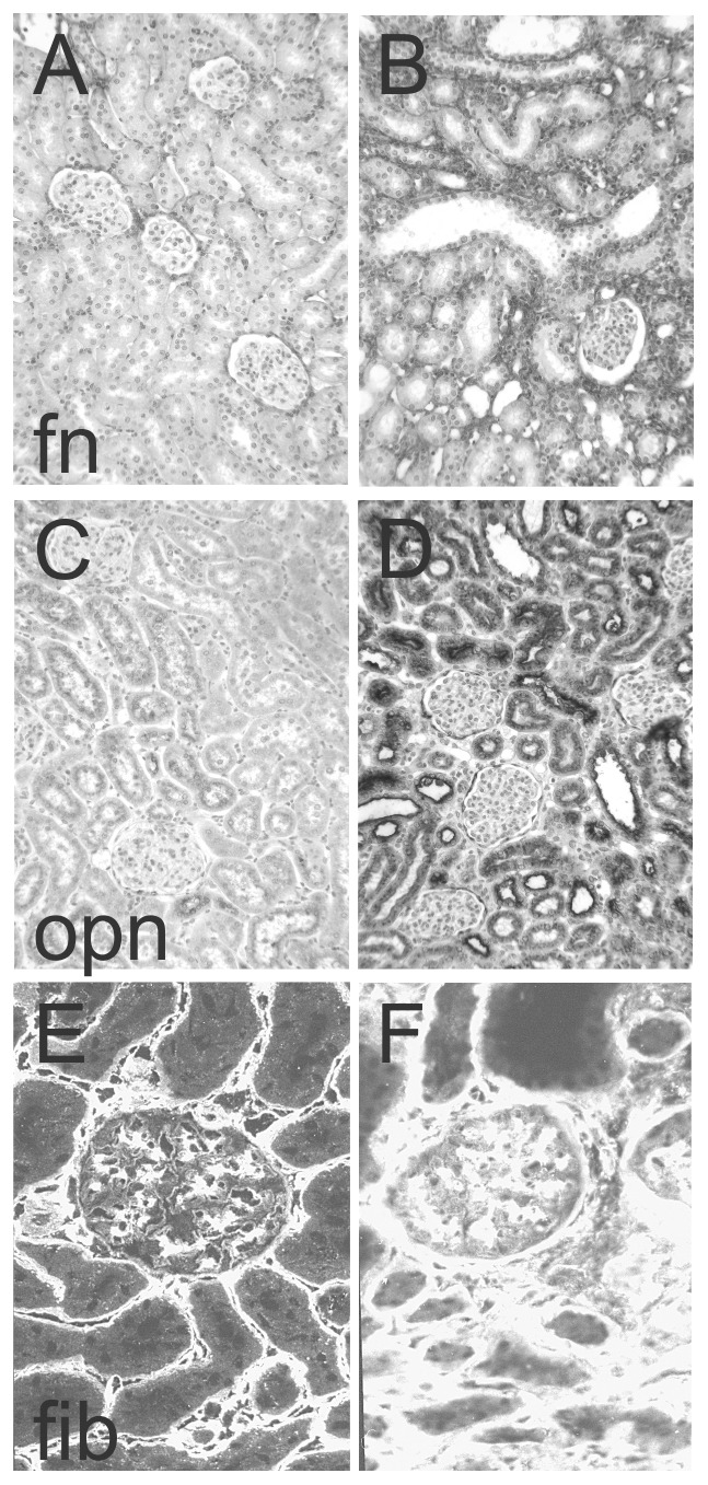
Figure 2. Exemplary photomicrographs of the immunohistochemical detection of A+B fibronectin (fn), C+D osteopontin (opn) and immunofluorescent detection of E+F fibrillin-1 (fib) in control kidneys (A, C, E) and in kidneys after unilateral ureteral obstruction (B, D, F).

Please note that A, B, C and D are enzymatic stainings which result in a dark stain, while E and F are fluorescent stainings resulting in a bright stain.
