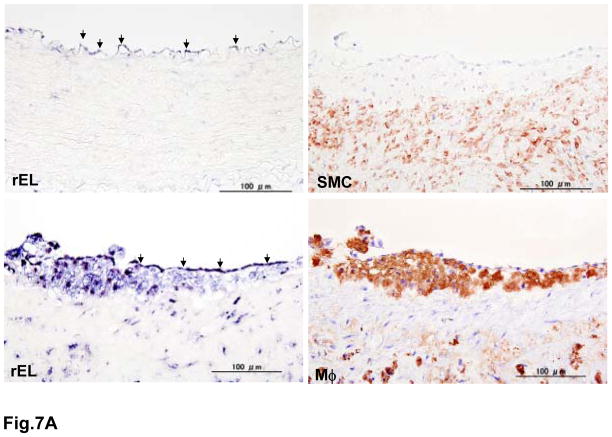
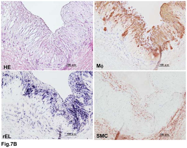
Fig. 7.
Demonstration of rEL expression in the endothelial cells and lesions of aortic atherosclerosis of WHHLMI rabbits (A). Serial sections were used for both in situ hybridization and immunohistochemical staining as described in the Materials and Methods. In normal aorta, weak expression of rEL (indicated by arrowheads) was noted in the endothelial cells (top, left). In the aortic lesions, both endothelial cells (indicated by arrowheads) and macrophages (M ) (bottom) showed rEL expression but only a few smooth muscle cells (SMC) showed rEL expression (top, right). In advanced lesions of WHHLMI rabbits (B), many macrophages and some smooth muscle cells showed rEL expression.


