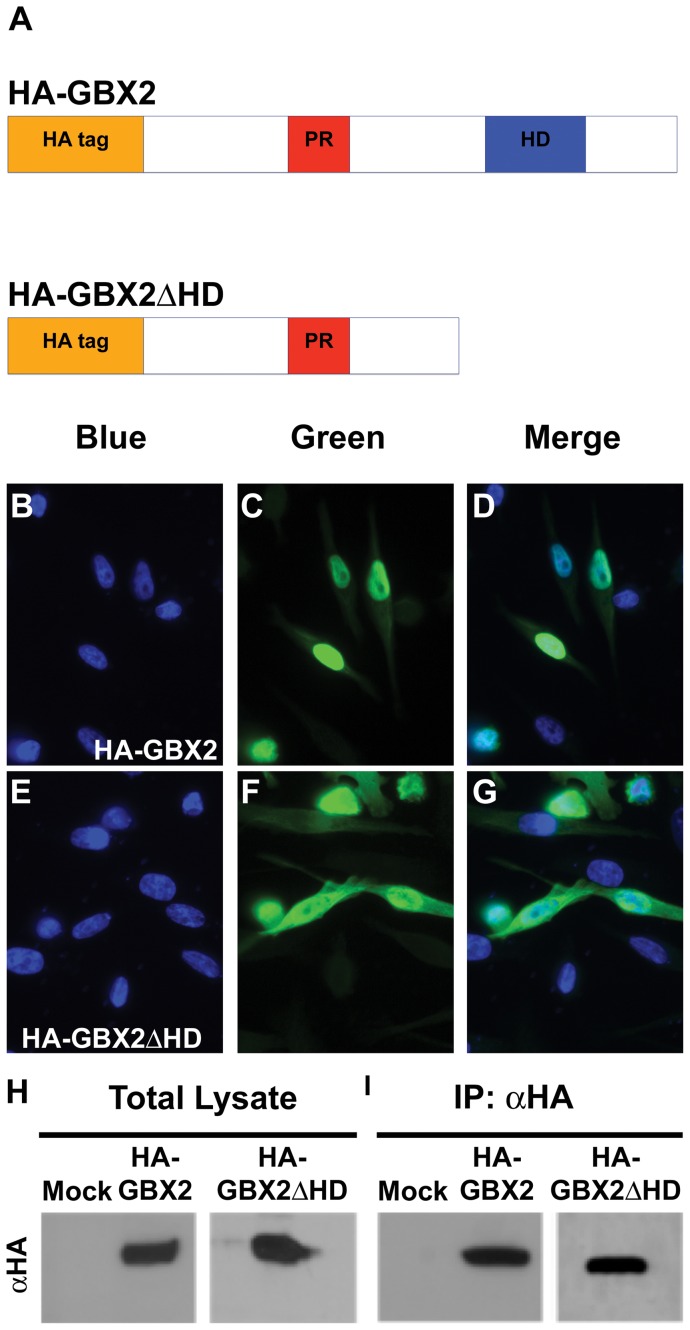
Figure 1. GBX2 overexpression and ChIP in human PC-3 cells.
(A) Schematic representation of the HA-GBX2 and HA-GBX2ΔHD recombinant proteins containing the proline-rich region (PR), DNA-binding homeodomain (HD), and the HA epitope tag located at the amino terminus. Immunoflouresence of transiently transfected human PC-3 cells with HA-GBX2 (C, D), and, HA-Gbx2ΔHD (F, G). Blue channel identifies DAPI staining in the nucleus (B, E). Green channel identifies GFP-GBX2 fusion proteins. (D, G) Merge displays nuclear localization of GFP-GBX2 fusion proteins. Western blots of total lysates (H) and HA-immunoprecipitated samples (I) from mock, HA-Gbx2, and HA-Gbx2ΔHD transfected PC-3 cells.

