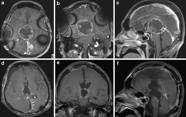
Fig. 2.

MR images taken during the course. a–c MRI with contrast during radiation showing enlargement of the tumor, indicating growing teratoma syndrome. d–f MRI with contrast after the third surgery showing a residual lesion on the right side of the third ventricle
