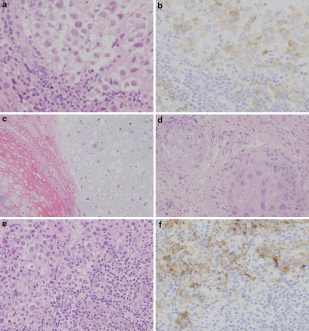
Fig. 4.

Photomicrographs of histological findings. a, b Initial biopsy specimen showing two-cell pattern of germinoma with large clear cells and small lymphoid elements. Immunohistochemical staining for PLAP revealed positive cells. (a H&E stain, b PLAP ×100). The specimens from the second surgery showed mature teratoma features with cartilage (c) and stratified squamous epithelium (d) (H&E stain ×200). e, f Biopsy specimen from retroperitoneal space lesion showing two-cell pattern of germinoma. Immunohistochemical staining for PLAP revealed positive tumor cells (e H&E stain, f PLAP ×200)
