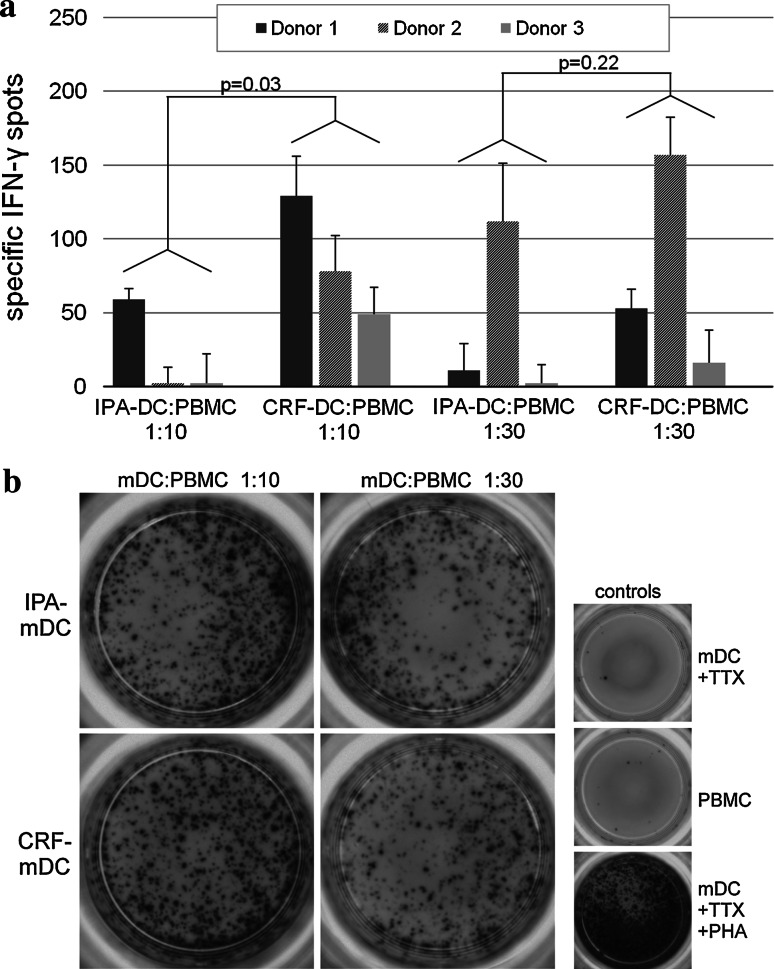
Fig. 6.
a Antigen-specific IFN-γ release of autologous PBMC after coculture with different tetanus toxoid- (TTX) loaded mDC (n = 3 different donors). Before determination of IFN-γ release in ELISPOT assay, TTX-loaded mDC were cultured with autologous PBMC for 1 week to allow expansion of antigen-specific T cells. Two different ratios were used as follows: 2 × 10⁶ PBMC+ 2 × 10⁵ mDC (ratio 10:1) and 2 × 10⁶ PBMC+ 6.6 × 10⁴ mDC (ratio 30:1). For IFN-γ ELISPOT, 5 × 10⁴ non-adherent cells after two rounds of stimulation were incubated in triplicates with 5 × 10⁴ TTX-loaded autologous PBMC for 48 h. Analysis of spots was performed by a computer-assisted video imaging analysis system. ELISPOT plates were analyzed in one single pass, and no spots were added or removed manually. TTX-specific spots were calculated by subtracting spots induced by unloaded control mDC from spot numbers induced by TTX-loaded mDC. b Images of the different cell subsets of donor 1 in IFN-γ ELISPOTS used for computer-assisted spot analysis. All experiments were performed in triplicates

