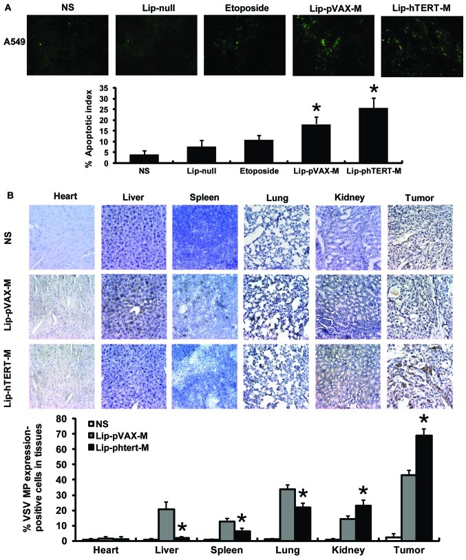
Figure 4.
Histochemical staining analysis. (A) Apoptotic cells within tumor tissues were evaluated by TUNEL assays. An apparent increase in the number of apoptotic cells and AI was observed within the tumor tissues in the Lip-phTERT-M and the Lip-pVAX-M groups compared with the other groups (*P<0.05). However, Lip-phTERT-M produced a greater increase. Data represent the mean AI ± SD of cancer cells. (B) Targeted antitumor effect analysis by immunohistochemical staining. The results indicated that Lip-phTERT-M restricted VSV MP overexpression to the tumor tissues rather than the other organs and had a superior specific antitumor effect (*P<0.05, compared with Lip-pVAX-M). AI, apoptotic index.

