Figure 2.
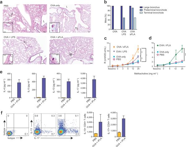
FLA promotes asthma-like responses to OVA. (a). Periodic acid-Schiff / Alcian blue staining of mucus-producing cells in the airway. Representative, low magnification (8×) images are shown (scale bar, 50 microns). Insets (40×) show expanded images of the indicated regions (scale bar, 10 microns). L, large airway; P, preterminal bronchioles. (b) Compiled data of mucus staining. n = 8 – 10 mice per group. (c,d) Mean values ± s.e.m. of airway resistance for intubated mice inhaling air (baseline), or aerosols of PBS containing the indicated concentrations of methacholine. n ≥ 8 mice/group. (e) Cytokine concentrations in cultures of lymph nodes excised from mice sensitized as indicated. IL-4 (left), IL-5 (middle) and IL-17 (right). (f) Intracellular staining for cytokines in pulmonary T cells. Shown are representative flow plots and bar histograms of mean ± s.e.m. numbers of CD4+ cells staining for IL-13 (left) and IL-17 (right).

