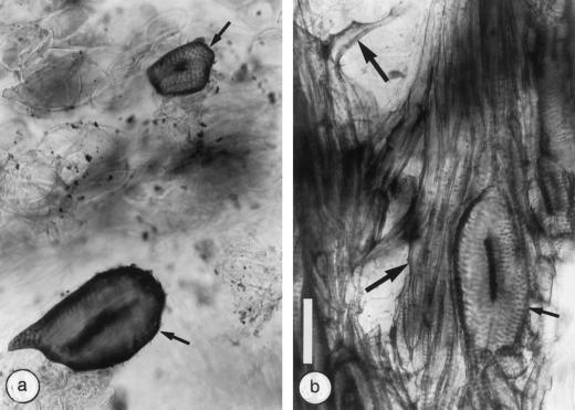
Figure 4.
Typical patterns of vascular tissues in 6-week-old A. tumefaciens-induced crown galls on tomato stems observed in thick, longitudinal radial sections cleared with lactic acid and stained with lacmoid. a, Circular vessels surrounded by parenchyma cells in wild-type stems. b, Circular vessels surrounded by fibers in the Nr mutant. Small arrows, Circular vessels; large arrows, fibers. Both photographs are at the same magnification (bar = 100 μm).

