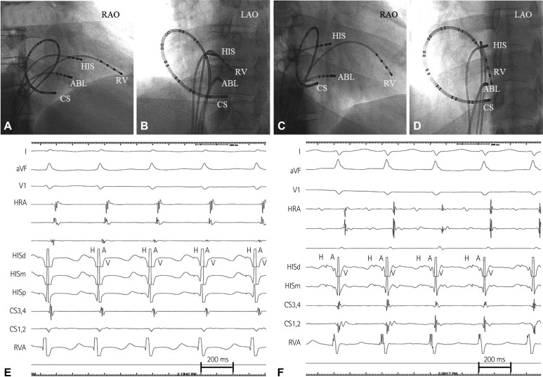
Fig. 1.
The mother's (A and B) and her son's (C and D) fluoroscopic images of endocardial mapping catheters in the right anterior oblique view (RAO) and the left anterior oblique view (LAO). The duodecapolar catheter was not fully engaged into the distal coronary sinus in both patients. The mother's (E) and her son's (F) intracardiac electrogram of typical atrioventricular nodal reentrant tachycardia (slow-fast type). During tachycardia, atrial activation is earliest at the HIS region and simultaneous with the ventricular activation. The VA interval (measured from the onset of ventricular activation on the surface electrocardiography to the earliest deflection of the atrial activation in the HIS electrogram) of both patients was <70 msec. HIS: His bundle, RV: right ventricle, HRA: high right atrium, CS: coronary sinus, ABL: ablation, VA: ventriculo-atrial, HISd: HIS distal, HISm: HIS middle, HISd: HIS distal.

