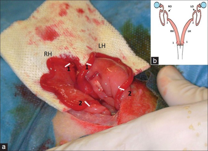
Figure 1.

(a) Schematic illustration of the rabbit uterus. Red marks: incisions sites (b) peroperative photograph of the two uterine horns. White marks: incisions sites (1) Proximal incisions (2) distal incisions. (RH) Right horn; (LH) Left horn; (RO) right oviduct t; (LO) Left oviduct
