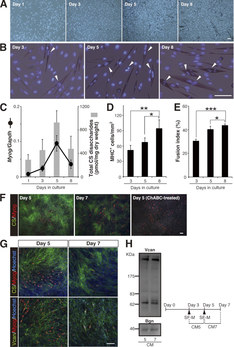
FIGURE 1.
Reduction in CS abundance correlates with the progression of multinuclear myotube formation in C2C12 myoblasts. A, temporal pattern of MHC immunostaining (brownish red) in parental C2C12 cells cultured in DM. Cell nuclei were stained with Hoechst 33342 (blue). Scale bar, 100 μm. B, higher magnification of the images in A. Arrowheads indicate MHC+ cells with two or more nuclei. Scale bar, 100 μm. C, changes in the levels of the Myog transcript (normalized to that of Gapdh) and of accumulated CS (estimated as the amount of total CS disaccharides) during myogenic differentiation of parental C2C12 cells (n = 10, Myog expression; n = 6, CS abundance; at each time point). Error bars represent S.D. D and E, the degree of myogenesis, including multinuclear myotube formation, was quantified based on MHC+ cell density (D) and on the fusion index of MHC+ cells (E) in parental C2C12 cell culture in DM at each time point (n = 4, for each time point; results are expressed as means ± S.D.; *, p < 0.05; **, p < 0.005; ***, p < 0.001). F–H, CS immunoreactivity was lower around Myog+ cells. Parental C2C12 cells on days 5 and 7 in DM culture were immunostained with anti-CS (green in F and G, upper panels), anti-versican core protein (Vcan, green in G, lower panels), and anti-Myog (red in F and G) antibodies. Cell nuclei were counterstained in blue with Hoechst 33342. Scale bar, 100 μm. The surrounding milieu of Myog+ cells, where versican core protein was uniformly distributed, was less immunoreactive for CS. ChABC treatment of the fixed cells before incubation with the anti-CS antibody eliminated the CS reactivity, confirming the specificity of the staining. H, temporal changes in the contents of CSPG core proteins in conditioned media (CM). Differentiating, parental C2C12 cells were incubated in serum-free medium (SF-M) for 48 h according to the time schedule shown at the bottom. Western blot analysis of CM5 and CM7 showed that expression levels of the core proteins, versican and biglycan (Bgn), are indistinguishable.

