FIGURE 2.
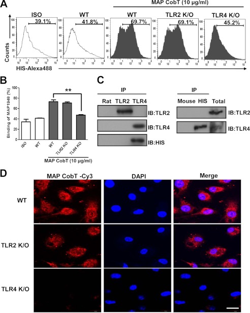
MAP CobT binds to TLR4, but not TLR2. A and B, bone marrow-derived DCs from WT, TLR2−/−, and TLR4−/− mice were treated for 1 h with MAP CobT (10 μg/ml) and stained with an Alexa 488-conjugated anti-His monoclonal antibody. The percentage of positive cells is shown in each panel. Bar graphs show the mean ± S.E. of the percentages of MAP CobT-Alexa 488 in CD11c+ cells from three independent experiments. Statistical significance (***, p < 0.001) is indicated for MAP CobT-treated TLR2−/− compared with MAP CobT-treated WT DCs. C, immunoprecipitation (IP) with His, TLR2, or TLR4 antibodies and immunoblotting (IB) with His, TLR2, or TLR4 antibodies. DCs were treated for 6 h with MAP CobT (10 μg/ml). Cells were harvested and cell lysates were immunoprecipitated with anti-rat IgG, anti-mouse IgG, anti-His, anti-TLR2, or anti-TLR4. Proteins were visualized by immunoblotting with His, TLR2, or TLR4 antibodies. Total cell lysate was used as an input control. D, fluorescence intensities of anti-MAP CobT bound to MAP CobT-treated DCs. DCs derived from WT, TLR2−/−, and TLR4−/− mice were treated for 1 h with MAP CobT (10 μg/ml), fixed, and stained with DAPI and a Cy3-conjugated anti-MAP CobT antibody (scale bar, 10 μm).

