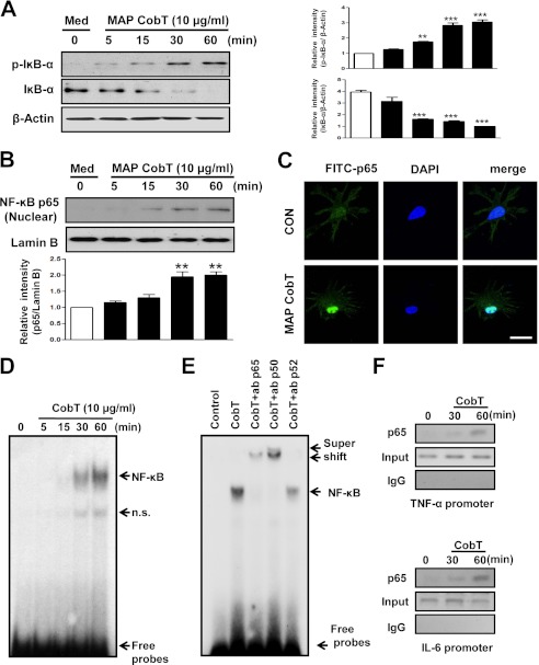
FIGURE 5.
DC maturation triggered by MAP CobT involves activation of NF-κB signal pathway. A and B, DCs were treated with 10 μg/ml of MAP CobT and protein expression is shown over time. Cells lysates or nuclear lysates were subjected to SDS-PAGE and immunoblot analyses were performed using specific antibodies to phospho-IκB-α (p-IκB-α), IκB-α (unphospho), and p65 NF-κB. β-Actin and lamin B were used as loading controls for cytosolic and nuclear fractions, respectively. Relative band intensity of each protein is expressed as a percentage compared with the value of untreated controls. The results shown are typical of three experiments for each condition. The data are shown as mean ± S.E. (n = 3) and statistical significance (*, p < 0.05; **, p < 0.01; or ***, p < 0.001) is indicated for treatments versus untreated DCs. C, effect of MAP CobT on the cellular localization of the p65 subunit of NF-κB in DCs. DCs were plated in covered glass chamber slides and treated for 1 h with MAP CobT. After stimulation, the p65 subunit in cells was evaluated by immunofluorescence as described under “Experimental Procedures” (scale bar, 10 μm). D, autoradiograph of EMSA performed with a 32P-labeled NF-κB nucleotide and nuclear extract from MAP CobT-treated DCs for the indicated time point. EMSA was performed as described under “Experimental Procedures.” n.s. indicates nonspecific band. E, supershift assay was performed using specific antibodies against NF-κB subunits, p65, p50, and p52 as described under “Experimental Procedures.” F, ChIP assays were performed on treated with MAP CobT using anti-p65 antibody for the indicated time point as described under “Experimental Procedures.”

